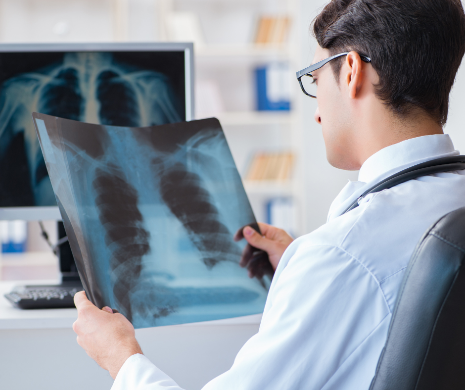
Diagnostic testing for lung cancer
Your family doctor or nurse practitioner may refer you to a specialist or order additional tests to check for lung cancer or other health problems. These tests may include a CT scan, a biopsy, a bronchoscopy or mediastinoscopy/mediastinotomy.
CT scan
If your chest x-ray shows anything abnormal or if your provider feels that they need more information, they will order a CT scan. A CT scan provides a more detailed image of the lungs and can show the size, shape and position of any lung tumors and can help find enlarged lymph nodes that might contain cancer that has spread.
Based on the outcome of your CT scan, you will be referred to a diagnostic specialist, typically a thoracic surgeon or respirologist, to provide a diagnosis and staging (how advanced your cancer is).
A thoracic surgeon specializes in surgeries of the chest (such as the lungs or esophagus). They also can diagnosis lung (and other) diseases of the chest and help determine the best treatment, surgical or otherwise. A respirologist is a doctor who specializes in the diagnosis and management of breathing conditions (including lung cancer, asthma and COPD).
Biopsy
You may be sent for a biopsy so that a piece of the tumor can be more closely examined. Depending on the location of the tumor, a tissue sample can be collected during a bronchoscopy or using a needle biopsy. If the tumor is difficult to reach or other methods do not work, your doctors may perform an open (or surgical) biopsy.
A needle biopsy is a method to remove a piece of lung tissue for examination. It may be required if imaging tests (x-ray, CT scan) cannot confirm that a nodule is benign, or a nodule cannot be reached by bronchoscopy or other methods. Needle biopsy is less invasive than surgical biopsy.
Bronchoscopy
Bronchoscopy is a test done using a bronchoscope, a thin, flexible tube that is inserted through your mouth or nose that has a small camera on the end to allow the operator to see deep into the lungs. Using special attachments to the end of the scope, it can be used to collect small tissue samples or to perform an ultrasound of the lymph nodes and other structures (an endobronchial ultrasound).
Mediastinotomy or mediastinoscopy
A mediastinotomy or mediastinoscopy is done to examine the space between the lungs, behind the sternum. It is done in an operating room at a hospital. This procedure is usually done as an outpatient procedure (you will not have to stay overnight at the hospital). The procedure will help your doctor to know if the cancer has spread to lymph nodes in the chest. The doctor may take tissue samples during the procedure.
PET scan
A PET (positron emission tomography) scan shows activity within cells. An injection of a sugar-based radioactive (tracer) solution is provided before the scan. Cells that are benign or consist of scar tissue are inactive and will not metabolize the sugar solution.
If other imaging tests don't provide a clear picture for diagnosis, a PET scan may be ordered. It may also be sued to see if lung cancer has spread or to find out how well cancer treatment is working. A PET scan can be combined with a CT scan using a PET/CT scanner. A combined scan will show the location, size and shape of any areas or growths that may be cancerous as well as a change in the cell metabolism that could indicate cancer.
This section was made possible by an unrestricted educational grant from Merck Canada, Sanofi Canada and Astra Zeneca Canada.
"I wasn't able to quit last time. I just can't do it."
Quitting smoking isn't easy — it often takes multiple attempts. Don't get discouraged. Each quit attempt is an opportunity to learn more about what tools and resources do and don't work for you and what triggers your cravings and how to deal with them.

